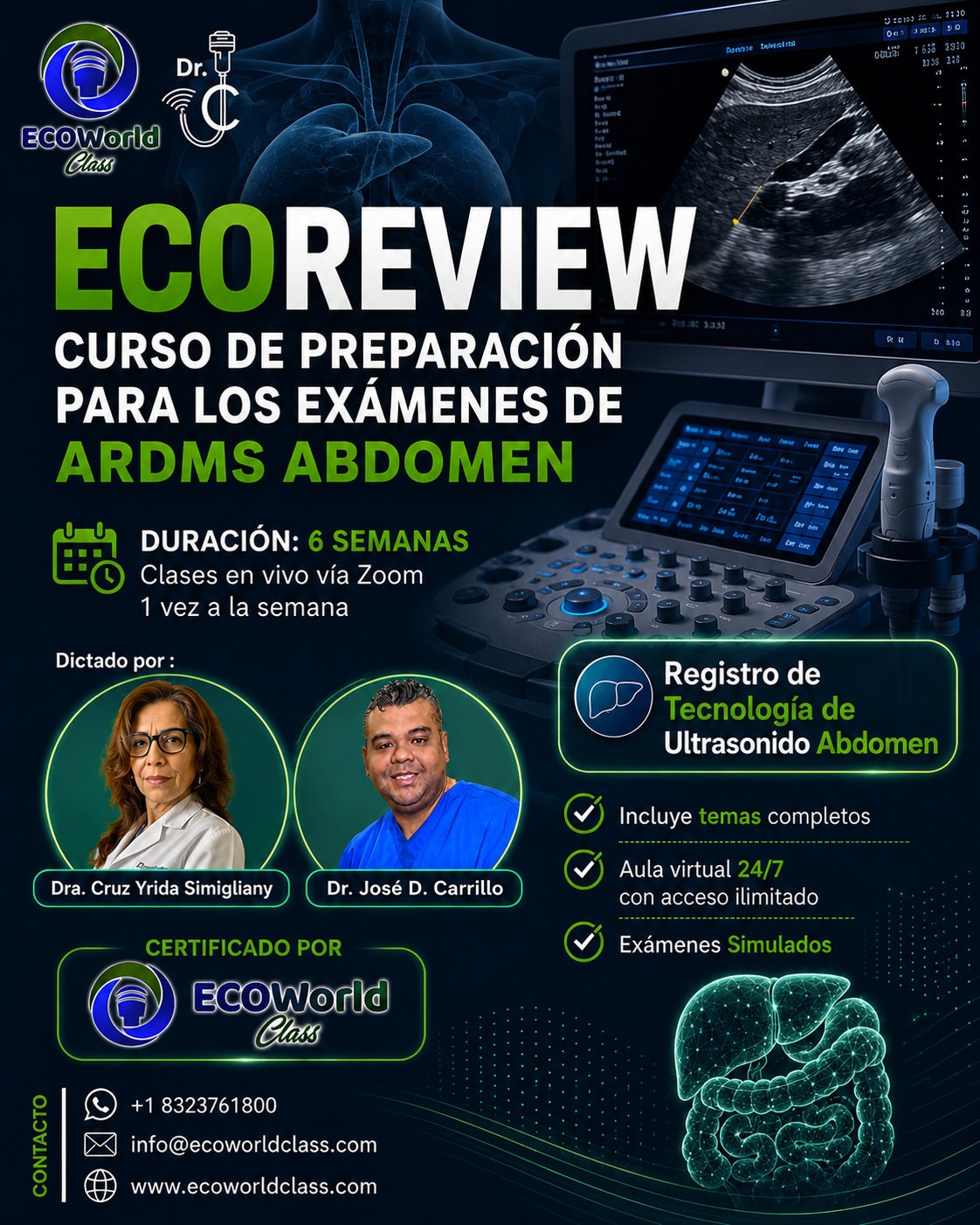
ECOREVIEW – ARDM Abdomen
Acerca de este curso


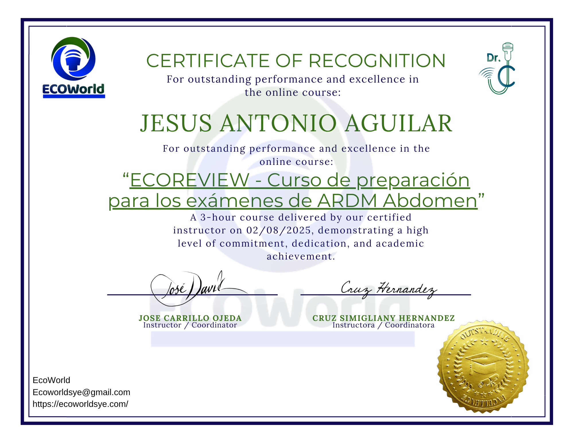
Certificado de finalización
Título del curso
-
Elemento de lista nº1
-
Elemento de lista nº2
-
Elemento de lista nº3
-
Elemento de lista nº4
-
Elemento de lista nº5
Modalidad de aprendizaje
Clases en video y recursos
Contenido en video con material de apoyo en pdf.
Acceso para siempre
Aprende a tu ritmo con un acceso de por vida al material.
Soporte de profesionales
Te ayudaremos en el proceso de aprendizaje respondiendo tus consultas.
Certificado de finalización
Aplica a un certificado al completar el 80% de las lecciones vistas.
Preguntas Frecuentes
A través de nuestra página web, botón de pago seguro o transferencia bancaria.
¿Qué métodos de pago están disponibles?
Aceptamos Zelle, PayPal, tarjetas de débito/crédito y transferencias bancarias.
¿Cuál es la modalidad del curso?
100% online, con acceso a clases grabadas y sesiones en vivo.
¿Necesito experiencia previa para tomarlo?
No, el curso está diseñado desde cero para cualquier profesional de salud con interés en aprender.
¿Puedo acceder al curso las veces que quiera?
Sí, tendrás acceso ilimitado a la plataforma durante toda la formación.
¿El curso me prepara para el examen ARDMS?
Sí, todo el contenido está enfocado en aprobar el examen SPI y los módulos clínicos.
¿Qué pasa si no puedo asistir en vivo?
Las clases quedan grabadas y disponibles en el aula virtual para que las veas en tu tiempo libre.
¿Obtengo certificado al finalizar?
Sí, recibirás un certificado digital de finalización avalado por nuestro equipo docente.
Necesito hablar ingles al 100%
No, te daremos todas las herramientas necesarias para que puedas aprobar el examen en inglés.
Contenido del curso
WELCOME
-
Bienvenidos al Curso de Ultrasonido Ecoreview Abdomen
01:20 -
EXPLICACION DE COMO MANEJAR EL AULA VIRTUAL
08:14 -
LIVECLASS ABD P1. WLC 24-02-26
01:17:06
WEEK 1
-
ABDOMEN ULTRASOUND PHYSICS
01:49:48 -
CHAPTER I
-
LIVER
01:53:07 -
LIVER CAPITULO II
01:14:06 -
CHAPTER II LIVER
-
practica evaluacion del lobe left hepatic
04:28 -
Practica Liver complete
05:21 -
Liveclass week I
02:07:46 -
live class 10/22/2025
01:46:04 -
LIVECLASS ABD P1 W1 03-03-26
01:38:55
WEEK 2
🟢 CHAPTER 3: NORMAL AND PATHOLOGICAL GALLBLADDER
🔬 Normal Anatomy
The gallbladder is a pear-shaped or saccular organ located in the gallbladder fossa, situated between the right hepatic lobe and the quadrate lobe of the liver. It consists of three parts: the fundus, body, and neck. Its main function is to store and concentrate bile, releasing it after meals to aid in digestion. In fasting patients, the gallbladder is typically distended and easier to evaluate.
Its normal length ranges from 7 to 10 cm, and the wall should measure ≤ 3 mm. An anechoic lumen without internal echoes is considered normal. The gallbladder receives blood supply from the cystic artery, which branches off the right hepatic artery. Normally, the cystic duct is not visualized unless dilated.
📏 Key Measurements
A wall thickness greater than 3 mm raises suspicion for inflammation. A gallbladder with no internal content and thin walls is considered normal. Any visible cystic duct dilation should be evaluated further, as it is not usually seen unless abnormal.
🟡 Cholelithiasis (Gallstones)
Gallstones appear as echogenic foci with posterior acoustic shadowing. They usually move when the patient is repositioned. Gallstones are the most common cause of gallbladder disease and may lead to obstruction and cholecystitis.
🟤 Biliary Sludge
Biliary sludge consists of low-level echoes that do not produce shadowing. It tends to layer dependently and shifts slowly with patient movement. Sludge is associated with bile stasis and may precede gallstone formation.
🌱 Gallbladder Polyps
Polyps are non-shadowing, non-mobile echogenic masses attached to the wall. They are usually benign, especially if smaller than 10 mm. If larger or rapidly growing, further evaluation is required due to the risk of malignancy.
🌀 Adenomyomatosis
Adenomyomatosis is a benign condition involving focal or diffuse wall thickening and intramural diverticula known as Rokitansky-Aschoff sinuses. It produces characteristic comet tail artifacts or ring-down artifacts, often in the gallbladder body.
🔥 Acute Cholecystitis
This condition is most often caused by cystic duct obstruction due to a gallstone. Sonographic findings include gallbladder wall thickening (>3 mm), positive sonographic Murphy's sign, pericholecystic fluid, and gallbladder distention. Shadowing gallstones and sludge are often present.
⚠️ Acalculous Cholecystitis
This is an inflammation of the gallbladder without gallstones, typically seen in critically ill or hospitalized patients. Diagnosis can be challenging, but ultrasound may show wall thickening, distention, and fluid, even in the absence of calculi.
💨 Emphysematous Cholecystitis
This life-threatening form of cholecystitis is caused by gas-producing bacteria. It presents with echogenic foci in the wall or lumen with dirty shadowing and reverberation artifacts due to gas. It carries a high risk of perforation and requires urgent intervention.
🧟 Gangrenous Cholecystitis
A complication of acute cholecystitis, gangrenous cholecystitis involves necrosis of the gallbladder wall. Findings include irregular wall thickening, sloughed membranes, intraluminal debris, and absence of Murphy's sign. It is associated with a higher risk of perforation.
🎗️ Gallbladder Carcinoma
Gallbladder cancer is rare but highly aggressive. Sonographic signs include a mass replacing the gallbladder, focal wall thickening, or a large non-mobile polypoid lesion. It is often associated with porcelain gallbladder and gallstones.
🔵 CHAPTER 4: BILIARY TREE (CBD and Intrahepatic Ducts)
🧠 Anatomy and Physiology
The common bile duct (CBD) is formed by the junction of the cystic duct and the common hepatic duct. It courses posterior to the pancreatic head and drains into the duodenum at the ampulla of Vater. On ultrasound, the CBD is typically visualized anterior to the portal vein in the portal triad.
The normal diameter of the CBD is up to 6 mm in adults under 60 years. One may add 1 mm per decade of life after age 60. In post-cholecystectomy patients, a CBD measuring up to 10 mm may still be considered normal.
🪨 Choledocholithiasis
Stones within the bile duct appear as echogenic foci that may or may not cast shadowing. They are usually associated with dilated intra- and extrahepatic ducts. Symptoms include jaundice, pain, and fever (Charcot’s triad in cholangitis).
🧬 Cholangitis
Cholangitis is an infection of the bile ducts, often caused by obstruction (e.g., from a stone or tumor). Ultrasound may show dilated ducts, thickened duct walls, and biliary debris. Clinical signs include fever, jaundice, and RUQ pain.
🎗️ Cholangiocarcinoma
This is a malignant tumor of the bile ducts, commonly at the hepatic duct bifurcation (Klatskin tumor). Sonographically, it appears as a mass at the hilum with proximal ductal dilation and no visible distal obstruction.
🛠️ Iatrogenic Injury / Strictures
Following surgery (e.g., cholecystectomy), the bile ducts may develop strictures or leaks. Ultrasound can reveal focal ductal dilation or fluid collections, and further imaging (e.g., MRCP or HIDA scan) is often required.
🪱 Biliary Parasites (Ascaris)
Parasitic infections such as Ascaris lumbricoides may appear as mobile tubular structures within the ducts. They are hypoechoic with echogenic borders and may move on real-time imaging.
🧪 Important Signs and Artifacts
Double Barrel Sign: The CBD and portal vein appear as two parallel anechoic tubes in the portal triad.
Shotgun Sign: Refers to dilated intrahepatic ducts running parallel to portal veins.
WES Sign (Wall-Echo-Shadow): Classic appearance of a stone-filled gallbladder; gallbladder wall, echogenic line from stones, and posterior shadowing — no visible lumen.
✅ Key RDMS Exam Takeaways
A CBD measuring >6 mm is considered abnormal, unless the patient is elderly or has had a cholecystectomy. Use color Doppler to differentiate ducts from vessels. A positive Murphy's sign with wall thickening is highly suggestive of acute cholecystitis. A non-shadowing, non-mobile lesion should raise concern for a polyp or malignancy. If the bile ducts are dilated without a visible cause, further imaging such as CT, MRCP, or ERCP is recommended.
-
Chapter 3
02:16:57 -
QUIZ GALLBLADDER
-
Chapter 4
01:14:47 -
QUIZ THE BILE DUCTS
-
PRACTICA LIVER COMPLETE + GALLBLADDER
-
LIVE CLASS 06/18/2025
01:57:46 -
PRACTICA OPTIMIZAR UNA IMAGEN
16:09 -
GALLBLADDER PROTOCOL
-
LIVE CLASS 04/13/2025
02:05:46 -
LIVE CLASS 28-10 WEEK 2
02:12:12 -
LIVECLASS ABD P1 W2 10-03-26
02:12:45
WEEK 3
🎓 Hello colleagues, welcome to a new study week in the ECOREVIEW RDMS-Abdomen course.
🗓️ This week we will be covering three key topics for the certification exam:
⸻
🔷 1. Pancreas
The pancreas is a retroperitoneal organ with both endocrine (insulin, glucagon) and exocrine (digestive enzymes) functions.
🧠 Essential exam points:
Echogenicity: Typically hypoechoic or isoechoic compared to the liver.
Common pathologies:
Acute pancreatitis: Enlarged, hypoechoic pancreas with possible fluid collections.
Chronic pancreatitis: Small, echogenic pancreas with calcifications.
Neoplasms: Adenocarcinoma (commonly in the head), associated with dilated pancreatic duct (Wirsung).
Pancreatic duct (Wirsung): Normally 13 mm
Reversed (hepatofugal) portal flow
Collateral circulation: Dilated gastric, splenic, or paraumbilical veins.
Splenomegaly: An indirect sign.
Ascites may also be present.
⸻
📌 Remember: These entities are highly tested in the RDMS-Abdomen exam. Be prepared to identify their ultrasound characteristics, correlate them with clinical findings, and apply diagnostic criteria.
See you in class to review images, exam-style questions, and clinical correlations. Let’s give it our all this week! 💪📚
-
PANCREAS
01:10:29 -
PANCREAS
-
SPLEEN
51:36 -
SPLEEN
-
AORTA ABDOMINAL
-
VIDEO PRACTICO I
12:39 -
live class 08/20/2025
01:50:45 -
Class live 11/04/2025
02:18:58 -
LIVECLASS ABD P1. W3. 17-03-26
02:05:30
WEEK 4
📋 Ultrasound Overview: Abdominal Aorta, Kidneys, and Adrenal Glands (with Emojis)
1. Abdominal Aorta 🏥
📐 Anatomy & Segments: Infradiaphragmatic portion from diaphragm to iliac bifurcation.
📏 Normal Diameter: 3 cm dilation)
Dissection (echogenic intimal flap)
Mural thrombus (echogenic wall adherent clot)
2. Kidneys 🩺
📏 Normal Measurements:
Length 9–12 cm; cortical thickness > 1 cm
Cortex is more echogenic than medulla
🔍 Technique:
Patient in lateral decubitus
3–5 MHz convex probe in coronal & transverse planes
💧 Common Findings:
Hydronephrosis: dilated pelvicalyceal system
Nephrolithiasis: echogenic stones with shadowing
Simple cysts: anechoic, thin walls, posterior enhancement
Solid masses: heterogeneous echoes, abnormal Doppler flow
📈 Doppler Parameters:
Interlobar artery PSV 0.7 suggests stenosis)
3. Adrenal Glands 🌟
📍 Location & Shape: Superior–medial to renal poles; triangular on right, semilunar on left.
🔍 Technique:
5 MHz convex probe
Oblique coronal plane over kidney
Contralateral lateral decubitus
🎨 Normal Appearance:
Length 2–4 cm; thickness < 1 cm
Hypoechoic cortex with echogenic medulla
⚠️ Pathologies to Recognize:
Adenoma: small, homogenous, stable nodule
Pheochromocytoma/Carcinoma: heterogeneous, hypervascular mass
Metastases: multiple lesions, rapid growth
Hyperplasia: diffuse bilateral enlargement
🔑 ARDMS/RVT Exam Tips:
Memorize normal size ranges and probe settings.
Use Doppler to assess renal artery velocities and resistive indices.
Identify and differentiate cystic vs. solid lesions and recognize vascular patterns.
Good luck on your exam! 🚀
-
ABDOMINAL VASCULATURE
01:17:34 -
ABDOMINAL VASCULATURE
-
THE URINARY TRACT
01:47:44 -
THE URINARY PART II
01:21:53 -
THE URINARY TRACT
-
ANDRENAL GLANDS
46:43 -
ADRENAL GLANDS
-
CLASS IN LIVE
02:33:42 -
LIVECLASSWEEK4 11-11-25
02:00:59 -
LIVECLASS AB P1 W4 24-23-26
WEEK 5
1. Gastrointestinal Tract and Abdominal Wall
Anatomy & Scan Approach
Segments: distal esophagus, stomach, duodenum, jejunum, ileum, colon (ascending, transverse, descending, sigmoid) and rectum.
Abdominal wall: rectus abdominis, external and internal obliques, transversus abdominis; linea alba and semilunar lines.
Scanning windows: longitudinal and transverse planes using intercostal, subcostal, and transabdominal approaches.
Normal Sonographic Appearance
Hollow-viscus walls: five-layer “onion‐ring” pattern (alternating echogenic and hypoechoic bands).
Luminal content: anechoic fluid or mixed echogenicity if gas is present, often producing reverberation or shadowing artifacts.
Abdominal wall musculature: hypoechoic muscle fibers with echogenic connective‐tissue striations.
Key Pathologies
Appendicitis: noncompressible appendix > 6 mm diameter, thickened wall, target sign on cross-section.
Bowel Obstruction: dilated loops (> 2.5–3 cm small bowel, > 6 cm colon), hyperactive (early) or reduced (late) peristalsis, air-fluid levels.
Diverticulitis: focal wall thickening, inflamed diverticulum often with echogenic gas, adjacent fat stranding.
Wall Hernias: fascial defect with protruding bowel or omentum; assess reducibility and vascular flow to exclude strangulation.
2. Thyroid
Anatomy & Technique
Right and left lobes connected by the isthmus; scan in longitudinal and transverse planes.
High-frequency linear transducer (10–15 MHz) for optimal resolution.
Normal Sonographic Appearance
Homogeneous, moderately echogenic parenchyma compared with strap muscles.
Uniform central vascularity on color Doppler.
Common Pathologies
Thyroid Nodules: stratify by echogenicity, margins, calcifications, and composition (TIRADS scoring).
Hashimoto’s Thyroiditis: heterogeneous, hypoechoic parenchyma with micronodules and reduced vascularity.
Graves’ Disease: diffusely enlarged gland with marked hypervascularity (“thyroid inferno”).
Multinodular Goiter: multiple nodules of varying sonographic appearance.
3. Non-Cardiac Chest
Scan Areas & Technique
Pleura, pleural space, accessible lung parenchyma (“comet-tail” zones), and upper mediastinum.
Use linear or curved array transducer in intercostal windows; patient seated or supine.
Normal Sonographic Signs
“Seashore sign” on M-mode: granular pattern below the pleural line.
A-lines: horizontal reverberation artifacts indicating aerated lung.
Pathologies & Artifacts
Pleural Effusion: anechoic or hypoechoic fluid collection above diaphragm; may show swirling echoes if exudative.
Pneumothorax: absence of lung sliding, presence of “barcode” or “stratosphere” sign on M-mode, absence of B-lines.
Consolidation: hypoechoic tissue with dynamic air bronchograms (echogenic punctate or linear “air” echoes).
Pulmonary Edema: multiple vertical B-lines (“comet tails”) that obliterate A-lines.
4. Retroperitoneum
Anatomy & Compartments
Spaces: suprarenal, perirenal (within Gerota’s fascia), pararenal anterior and posterior.
Major structures: kidneys, proximal ureters, adrenal glands, and head of pancreas (retroperitoneal portion).
Scanning Technique
Subcostal and paravertebral windows; image in both long and short axes.
Normal Sonographic Appearance
Kidney: cortex is mildly hyperechoic relative to liver/spleen, medulla is hypoechoic, and the sinus is echogenic.
Adrenal Gland: thin triangular or crescent shape with echogenicity similar to surrounding fat.
Key Pathologies
Hydronephrosis: dilation of the renal pelvis and calyces, graded I–IV by degree of pelvicalyceal dilation and cortical thinning.
Renal Masses: simple cysts are anechoic with posterior acoustic enhancement; solid masses are heterogeneous, often with internal vascularity.
Adrenal Lesions: adenomas (homogeneous, slightly hyperechoic), metastases or pheochromocytomas (heterogeneous, hypervascular).
Acute Pancreatitis: peripancreatic fluid collections or edematous hypoechoic areas around the pancreatic head.
-
THE GASTROINTESTINAL TRACT AND ABDOMINAL WALL
01:13:29 -
THE GASTROINTESTINAL TRACT AND ABDOMINAL WALL
-
NONCARDIAC CHEST AND RETROPERITONEUM , THE FACE AND NECK
01:51:14 -
NONCARDIAC CHEST AND RETROPERITONEUM
-
LIVECLASS WEEK5 18-11-25
01:29:46 -
LIVECLASS P1 W5 ABD 31 03 26
02:04:22
WEEK 6
-
THE MALE PELVIC
02:27:37 -
SKELETAL MUSCLE HIP AND BREAST
01:32:42 -
THE MALE PELVIC
-
CLASE 16/07/2025
01:52:27 -
SKELETAL MUSCLE HIP AND BREAST
-
LIVE CLASS Q&A
02:18:07 -
protocolo Thyroid
34:48 -
LIVECLASS 25-11-25
01:56:47 -
LIVECLASS 02-12-25
02:10:24 -
LIVECLASS 09-12-25
02:04:20 -
LIVECLASS ABD P1 W6 07-04-26
02:14:53
EXAMEN REPORT
Hello, dear students! 🌟 Welcome to this module, thoughtfully designed to help you master the art of writing clear, concise, and professional ultrasound reports before sending them on to the radiologist. 📋✨ Here, you’ll learn how to highlight the most important findings, record accurate measurements, and draw appropriate conclusions—all in a way that showcases your technical expertise.
Throughout this module, you’ll practice:
• Describing organ size and echotexture with precision 🔍
• Noting the presence or absence of masses, cysts, stones, or sludge 🏥
• Measuring structures like the gallbladder wall and common bile duct, and interpreting those values 📏
• Reporting on vascular flow patterns, such as hepatopetal portal vein flow 🌊
By the end of this exercise, you’ll be able to confidently compose a complete, well-structured report that reads smoothly and provides the radiologist with all the data they need. Let’s get started—your future self (and your patients!) will thank you. 🙌🏼😁
Example : The liver measures 13.2 cm and demonstrates a homogeneous echotexture without evidence of intrahepatic masses or cysts; increased echogenicity from fatty infiltration consistent with Grade I steatosis may be present. The gallbladder wall is thin at 0.30 cm, the lumen is anechoic, and no gallstones or sludge are identified. The common bile duct measures 0.78 cm in diameter, within normal limits, and shows no dilatation. The portal vein exhibits hepatopetal flow with a diameter of 0.93 cm, also within expected parameters. All findings are consistent with a normal ultrasound examination of the liver, gallbladder, and biliary tree.
-
1 REPORT
-
REPORT II
-
REPORT III
EVALUATION ARDMS
Estimados estudiantes, atención:
– La Evaluación Nº 1 constará de 70 preguntas de estilo RDMS.
– Dispondrán de 1 minuto para responder cada pregunta.
– No habrá reintentos: una vez enviado el examen, no podrán repetir ninguna pregunta.
Les recomiendo gestionar bien su tiempo: si una pregunta les lleva más de un minuto, continúen avanzando y regresen a ella —en la medida de lo posible— dentro del tiempo asignado. ¡Éxito en su preparación!
-
QUESTION TYPE RDMS
-
ARDMS-ABDOMEN 1
-
EXAMEN RDMS CV FORM
VIDEOS PRACTICOS
-
CONOCIENDO EL EQUIPO
18:16 -
MODO DOPPLER COLOR
08:22 -
MODO B
05:37 -
MODO DOPPLER ESPECTRAL
12:46
EXAM FORMS
-
EXAM FORM ADM