-
LevelIntermedio
-
CertificadoCertificado de finalización
A course by
Tags
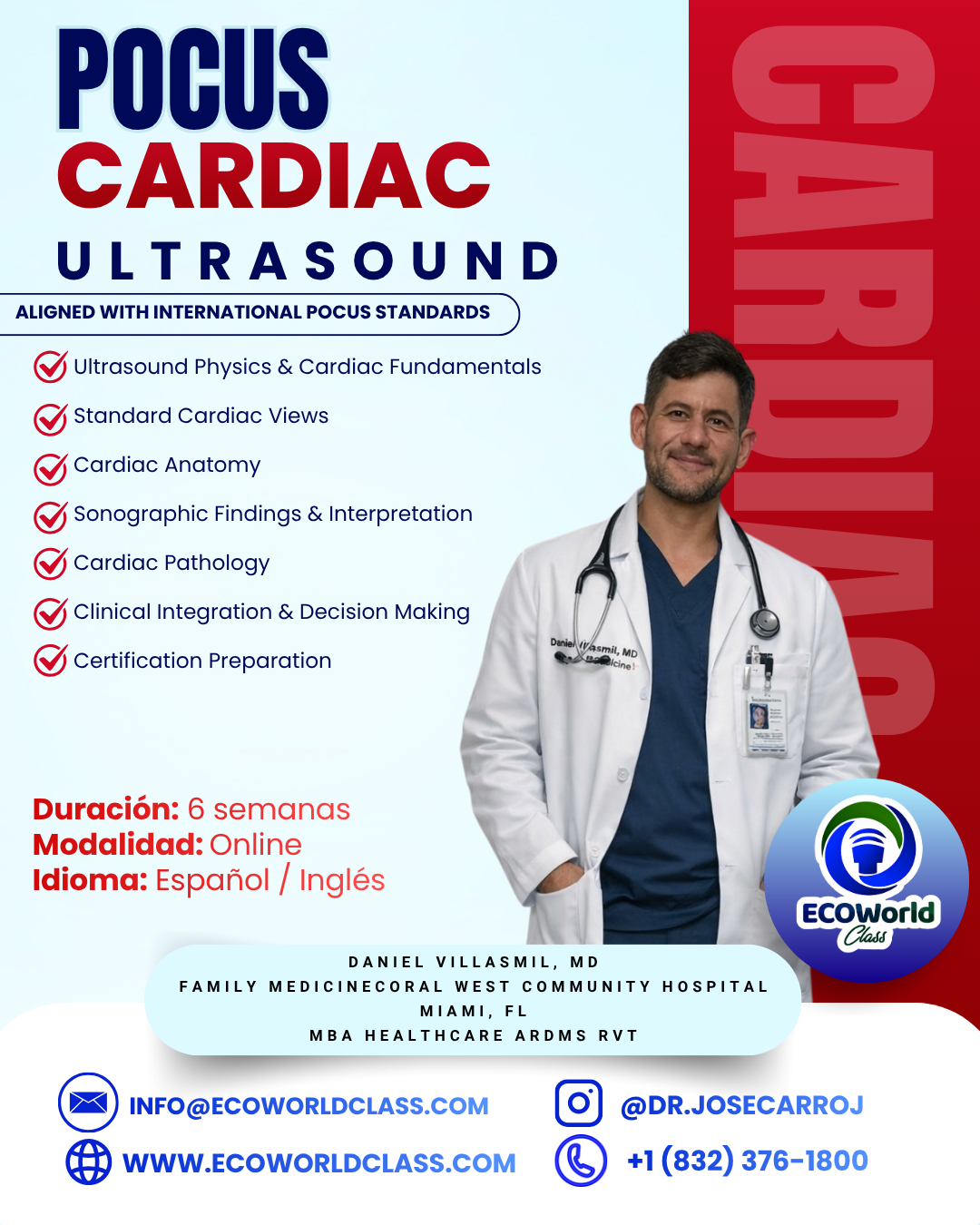
• Course Title: POCUS Cardiac Ultrasound Program
• Duration: 6 Weeks (Total: 40 Hours)
• Format: Online (Live + Recorded) + Optional Hands-on
• Language: English / Spanish
• Level: Beginner to Intermediate (Clinically Oriented)
• Instructor: Dr. José David Carrillo, MD – ARDMS (RVT, RDMS OB/GYN, SPI)
This program provides a comprehensive and clinically oriented approach to cardiac point-of-care ultrasound (POCUS). The course is designed to develop competency in image acquisition, anatomical recognition, sonographic interpretation, and clinical decision-making. The curriculum is aligned with the standards and assessment structure of the POCUS Certification Academy, preparing learners for Clinical Case Assessments, Peer Evaluation, and Clinical Video Submission.
Program Director and Clinical Content Lead:
Dr. José David Carrillo, MD
• Master’s Degree in Clinical Echocardiography
• Diploma in Diagnostic Ultrasound
• ARDMS Certified:
o Registered Vascular Technologist (RVT)
o RDMS – Obstetrics and Gynecology
Dr. Carrillo is responsible for curriculum design, academic oversight, clinical accuracy, and
competency validation. The program is developed under his supervision to ensure alignment
with evidence-based practice and international ultrasound education standards.
By the end of this program, participants will be able to:
• Perform standard cardiac POCUS views (PLAX, PSAX, Apical, Subcostal)
• Identify normal and abnormal cardiac anatomy
• Recognize key sonographic patterns and artifacts
• Assess ventricular function and hemodynamic states
• Identify common cardiac pathologies using POCUS
• Integrate ultrasound findings into clinical decision-making
• Prepare for POCUS Certification pathways
Module 1: Ultrasound Physics & Cardiac Fundamentals
• Basic ultrasound physics applied to cardiac imaging
• Transducer selection and orientation
• Image optimization (gain, depth, focus)
• Common artifacts
Module 2: Standard Cardiac Views (12%)
• Parasternal Long Axis (PLAX)
• Parasternal Short Axis (PSAX)
• Apical 4-Chamber
• Subcostal view
• Indications and scanning protocols
Module 3: Cardiac Anatomy (35%)
• Cardiac chambers (LV, RV, LA, RA)
• Valves (mitral, aortic, tricuspid)
• Septum and pericardium
• Anatomy in B-mode, M-mode, and Doppler
Module 4: Sonographic Findings & Interpretation (47%)
• Pattern recognition
• Functional assessment (LV function, RV strain)
• Pericardial vs pleural effusion
• Artifact recognition
• Basic measurements
Module 5: Cardiac Pathology (6%)
• Pericardial effusion and tamponade
• Cardiomyopathy
• Right heart strain (PE suspicion)
• Hypovolemia vs fluid overload
• Acute valvular abnormalities
• Cardiac standstill
Module 6: Clinical Integration & Decision Making
• POCUS in shock (hypovolemic, cardiogenic, obstructive)
• Rapid bedside assessment
• Integration with clinical findings
Module 7: Certification Preparation
• Clinical case-based simulations
• Image interpretation exercises
• Diagnostic reasoning
• Exam-style questions
Participants will be evaluated using a system aligned with POCUS standards:
1. Clinical Case Assessment
• Scenario-based questions
• Image interpretation
• Clinical decision-making
2. Peer Evaluation Preparation
• Image quality
• Gain and depth optimization
• Proper probe positioning
• Acquisition of diagnostic views
3. Practical Component (Optional)
• Image acquisition practice
• Video case preparation (for certification readiness)
Upon completion, participants will be prepared to:
• Sit for the POCUS Fundamentals Certificate (if required)
• Advance to POCUS Cardiac Clinical Certification
• Complete Peer Evaluation and Clinical Case Assessment
• Video lectures (on-demand)
• Live interactive sessions
• Clinical case library
• Image interpretation bank
• Downloadable guides
• Physicians (MD)
• Nurses (RN)
• Physician Assistants (PA)
• Sonographers
• Healthcare professionals interested in POCUS
• Basic medical knowledge
• Access to ultrasound machine (recommended for practice)
• Commitment to clinical training
Early bird discount price: $700 USD
Payment methods available:
* 💳 Stripe
* 💻 PayPal
* 🏦 Zelle: info@ecoworldclass.com
has revolutionized how clinicians approach diagnostic challenges at the bedside. Rather than waiting for a formal imaging suite study, POCUS allows for immediate, real-time visualization to answer specific clinical questions
Currículum del curso
Module 1: Ultrasound Physics and Doppler Principles
-
HEMODYNAMIC Part I
02:03:41 -
HEMODYNAMIC Part II
01:57:25 -
DOPPLER SPECTRAL
02:15:44 -
DOPPLER COLOR
02:18:16 -
LIVECLASS RVT P1. W1. 23-01-26
02:48:00
Module 2: Carotid Ultrasound
-
ANATOMY PART I
01:21:08 -
EXTRACRANIAL CEREBROVASCULAR
02:33:51 -
ANATOMY
01:14:14 -
PATHOLOGIES
01:20:07 -
LIVECLASS RVT P1. W3. 06/02/2026
02:27:17
Module 3: Peripheral Arterial Ultrasound
-
ANATOMY 2DA PART
48:02 -
LOWER ARTERY EXTREMITY PART I
02:52:35 -
LOWER EXTREMITY II PART
02:11:28 -
LOWER EXTREMITY III PART
35:24 -
LIVECLASS RVT P1 W6 06-03-26
02:19:42 -
LIVECLASS RVT P1. W7. 13-03-26
01:35:04
Module 4: Venous Ultrasound (DVT and Insufficiency)
-
VASC CTL: Extremity Venous Pathology – Exam Conversion
-
Peripheral Venous
-
LOWER EXTREMYTY VENOUS PART I
02:18:18 -
LOWER EXTREMITY VENOUS II PART
-
Q & A WEEK VI
02:15:51 -
LIVECLASS RVT PI W7 II 20-03-26
00:00
Module 5: Abdominal Vascular Ultrasound
-
VASCULAR ABDOMINAL
01:36:41 -
ABDOMEN VASCULAR DRA CRUZ
01:17:33 -
ECOPILDORA DOPPLER ABDOMINAL
02:28:44 -
Q & A ABDOMEN
00:00 -
VASC CTL Abdomen & General Pathology – Exam Conversion
-
Abdominal Vascular Studies
module 6:
Final Exam for POCUS Certification
-
EXAM POCUS CERTIFICATION