0 (0 Ratings)
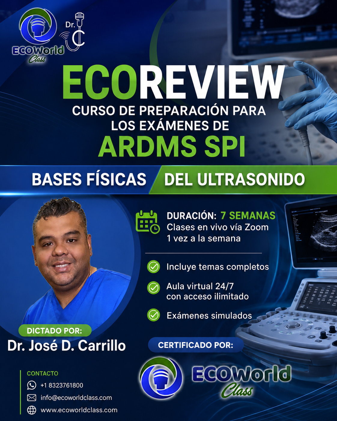
ECOREVIEW – Curso de preparación para los examenes de ARDMS SPI
$600.00
$900.00
-
LevelIntermedio
-
CertificadoCertificado de finalización
¡Hola, bienvenido de nuevo!
1. Comprender los principios físicos del ultrasonido y su interacción con los tejidos.
2. Analizar los modos de imagen y optimizar la resolución en cada aplicación.
3. Dominar la formación del haz, focalización y ajustes de compensación.
4. Evaluar bioefectos, bioseguridad e instrumentación.
5. Aplicar técnicas Doppler y reconocer artefactos.
6. Prepararse eficazmente para el examen SPI-ARDMS con simulacros y estrategias de
resolución rápida
 Dr. José David Carrillo
Médico General
• Ingeniero Industrial (2011)
• ARDMS RDMS-SPI, APCA Certified
• Fundador y Director de Ecoworld Class LLC
Plan de Estudios (6 Semanas)
🔹 Semana 1
Fundamentos del sonido: ondas, frecuencia, longitud de onda, impedancia.
Parámetros del ultrasonido e interacción con tejidos.
🔹 Semana 2
Modos de imagen: A-mode, B-mode, M-mode. Resolución axial vs. lateral. Tipos de
transductores y sus aplicaciones.
🔹 Semana 3
Alcance de imagen: profundidad, ganancia, compensaciones TGC. Formación del haz,
focos simples y múltiples, zoom y persistencia.
🔹 Semana 4
Bioefectos y seguridad: índices mecánico y térmico. Intensidades de pulso y continua.
Resolución temporal. Componentes de la consola e instrumentación.
🔹 Semana 5
Doppler y Artefactos (incluye 3 clases en vivo de 3 h c/u): Doppler color, PW, CW.
Principales artefactos y cómo evitarlos.
🔹 Semana 6
Repaso general: simulacros de examen, preguntas estilo SPI-ARDMS y técnicas de
resolución rápida de casos.
✔️ Fundamentos de acústica médica y parámetros de ultrasonido
✔️ Características y aplicaciones de los modos A, B y M
✔️ Técnicas de enfoque, barrido y formación de haz
✔️Seguridad, bioefectos e intensidades empleadas
✔️Principios y práctica de Doppler color, pulso y continuo
✔️Identificación y corrección de los artefactos más frecuentes
✔️ Estrategias de estudio y simulacros para aprobar el SPI-ARDMS
- Modalidad: 100% online
- Acceso a clases en video y material complementario
- Aprendizaje práctico y directo, centrado en el uso del ecógrafo
Instructor: Dr. José David Carrillo, médico e ingeniero especializado en ecografía clínica
¿Por Qué Elegir Este Curso?
• Experiencia comprobada: clases impartidas por un instructor certificado
• Formato mixto: teoría grabada + sesiones en vivo interactivas en la semana 5, 3 clases
en vivos.
• Material exclusivo: guías de estudio, resúmenes visuales y banco de preguntas.
• Enfoque práctico: simulacros con retroalimentación personalizada.
• Flexibilidad: acceso 24/7 a la plataforma y grabaciones de clases en vivo.
💵 Costo total: 600 USD
• Formas de pago disponibles:
💳 Stripe
💻 PayPal
🏦 Zelle → info@ecoworldclass.com
• ¿Tienes un cupón? Aplícalo al realizar el pago y obtén tu descuento.
 ¿Tienes dudas?
Contáctanos directamente vía WhatsApp:
Currículum del curso
WELCOME SONOGRAPHER
¡Bienvenidos al curso ECoReview SPI! 🎉👩⚕️👨⚕️
Estamos muy emocionados de que te unas a esta aventura de aprendizaje donde profundizaremos en los principios y la instrumentación del ultrasonido. Aquí descubrirás cómo el sonido interactúa con los tejidos, mejorarás tus habilidades técnicas y estarás listo para aprobar tu examen SPI con confianza.
🔹 Duración: 6 semanas intensivas
🔹 Modalidad: 100% online, con clases pregrabadas y sesiones en vivo
🔹 Recursos: PDFs, guías interactivas, exámenes de práctica
Prepárate para:
– Explorar las variables acústicas y su impacto en la calidad de imagen 🔊
– Dominar modos de imagen (B, M, Doppler) 📈
– Optimizar parámetros para diagnósticos precisos 🎯
¡Vamos a comenzar! 🚀
Pronto recibirás el enlace de acceso a la plataforma. ¡Nos vemos en clase! 👩🏫✨
-
WELCOME
01:10 -
Class live Welcome
01:17:30 -
LIVECLASS P2 WEEK WELCOME 06-04-26
01:21:43
WEEK 1
Key Physics Concepts for SPI (ARDMS) Exam
1. Basic Sound Properties
Frequency (f): Number of cycles per second (MHz in medical ultrasound).
Wavelength (λ): Distance a wave travels in one cycle; λ = c / f (c ≈ 1540 m/s in soft tissue).
Propagation Speed (c): Speed of sound in a medium (bone ≈ 4080 m/s, soft tissue ≈ 1540 m/s, lung ≈ 500 m/s).
Acoustic Impedance (Z): Z = ρ·c (density × speed); governs reflection at tissue interfaces.
2. Pulsed Ultrasound Parameters
Pulse Duration (PD): Time from the start to end of one pulse (PD = n × T, where n = cycles/pulse, T = period).
Pulse Repetition Period (PRP): Time from the start of one pulse to the start of the next (includes “listening” time).
Pulse Repetition Frequency (PRF): Number of pulses emitted per second (PRF = 1 / PRP).
Duty Factor (DF): Fraction of time the transducer is “on” transmitting (DF = PD / PRP).
Exam Tip: Low DF (< 1%) minimizes bioeffects but limits average power.
3. Intensity Definitions & Bioeffects
Spatial: How intensity varies across the beam (SP = single-point; SA = averaged across cross-section).
Temporal: How intensity varies over time (TP = peak; TA = averaged over pulse or PRP).
Key Intensities:
SPTP (Spatial‐Peak, Temporal‐Peak): Highest instantaneous intensity.
SPPA (Spatial‐Peak, Pulse‐Average): Peak intensity averaged over a pulse.
SPTA (Spatial‐Peak, Temporal‐Average): Most relevant for thermal bioeffects—averaged over PRP.
SATA (Spatial‐Average, Temporal‐Average): Overall beam intensity.
Regulatory Focus: Keep SPTA below FDA limits (< 720 mW/cm² for adult scans).
4. Sound–Medium Interactions
Reflection: At impedance mismatches;
– Specular: smooth interfaces (organ boundaries).
– Diffuse (Scattering): rough surfaces or small structures (RBC scattering → brightness).
Transmission & Refraction: Beam bends when crossing oblique interfaces with different speeds (Snell’s law).
Attenuation: Loss of beam intensity with depth;
– Causes: Absorption (heat), scattering, reflection.
– Coefficient (α): ≈ 0.5 dB/cm/MHz in soft tissue.
– Half‐Value Layer: Depth where intensity is halved (3 dB loss).
5. Clinical Relevance
Frequency vs. Resolution/Penetration: Higher f → better axial & lateral resolution (↑ detail) but ↑ attenuation (↓ depth).
Gain & TGC: Compensation for depth‐dependent attenuation; avoid over‐amplifying noise.
Axial Resolution: PD / 2; improved by shorter pulses (fewer cycles, wide bandwidth).
Lateral Resolution: Beam width at focus; improved by focusing, lower f, or smaller aperture.
Study Strategy:
Memorize formulas: λ = c/f, PRF = 1/PRP, DF = PD/PRP.
Know intensity categories and which relate to thermal (SPTA) vs. mechanical (MI) bioeffects.
Understand attenuation and its frequency‐dependence (α ∝ f).
Practice scenario questions on optimizing pulse parameters (e.g., adjusting PRP/PRF for deeper imaging).
Good luck on your SPI exam! 🚀
-
PHYSICAL PRINCIPLES OF THE SOUND
01:19:27 -
PHYSICAL PRINCIPLE
-
PULSE PARAMETERS IN ULTRASOUND
36:47 -
Pulse Parameters in Ultrasound
-
Interaction of Sound and Media
01:28:07 -
Interaction of Sound and Media
-
Sound Intensity in Ultrasound
39:13 -
Sound Intensity in Ultrasound
-
CONOCIENDO EL EQUIPO
18:16 -
EXAM RDMS
-
CLASS IN LIVE 09/09/2025
02:13:26 -
LIVECLASS P1. WEEK 1. 12-01-26
01:44:38 -
LIVECLASS SPI. P1. W1. 19-01-26
02:31:52 -
LIVECLASS SPI P2 W1 13-04-26
02:31:36
WEEK 2
1. Pulse-Echo Instrumentation
Pulser/Transmitter:
Generates electrical pulses that excite the piezoelectric crystal in the transducer.
Controls pulse amplitude and repetition rate (PRF).
Transducer:
Converts electrical pulses into ultrasound waves (transmission) and returning echoes into voltages (reception).
Houses matching layers and backing material to optimize bandwidth and damping.
Receiver/Signal Processing:
Preamplifier & TGC: Boosts returning echo voltages and applies depth-dependent gain.
Demodulator (Detector): Extracts the echo envelope (rectification + smoothing).
Scan Converter: Maps echo amplitude and time-of-flight into 2D image pixels (gray‐scale display).
Display & Storage:
Gray‐scale “B-mode” images with adjustable parameters (gain, dynamic range).
Options for M-mode, Doppler overlays, cine loops, and digital archiving.
2. The Range (Depth) Equation
where:
𝐷
D = depth of reflecting structure
𝑐
c = speed of sound in tissue (~1540 m/s)
𝑡
t = measured time from pulse emission to echo reception
Division by 2 accounts for the round-trip path.
Clinical Implications:
Maximum Imaging Depth: Limited by the pulse repetition period (PRP); PRF must be low enough to allow echoes from the deepest structure to return before the next pulse.
Axial Resolution: Depends on pulse duration and bandwidth; shorter pulses → better resolution.
Time‐to‐Distance Calibration: The machine uses the range equation in real time to convert each echo’s time delay into a pixel depth on screen.
3. Harmonic Imaging
Principle:
As ultrasound propagates, nonlinear tissue responses generate harmonic frequencies (multiples of the transmitted fundamental frequency).
Implementation:
Tissue Harmonics: Transmit at fundamental frequency (f), receive at 2f.
Pulse-Inversion or Power-Modulation Techniques: Cancel fundamental echoes and isolate harmonic signals for display.
Advantages:
Improved Lateral Resolution: Harmonic beam is narrower.
Reduced Artifacts: Less near-field noise, fewer side-lobe and reverberation artifacts.
Enhanced Contrast: Tissue harmonics arise mainly along the beam axis, improving image clarity in difficult-to‐image patients.
-
Pulsed Echo Instrumentation
44:24 -
Pulsed Echo Instrumentation
-
Rangue Equation
23:03 -
Rangue Equation
-
Harmonic
21:08 -
Harmonic
-
ARDMS WEEK 2
-
PRACTICE WEEK 2
11:46 -
class in live
01:23:55 -
LIVE CLASS 27-10 WEEK2
01:54:51 -
LIVECLASS SPI P1. W2. 26-01-26
02:38:45 -
LIVECLASS SPI P2 W2 20-04-26
02:05:23
WEEK 3
-
ULTRASOUND TRANSDUCER
47:52 -
ULTRASOUND TRANSDUCER
-
SOUND BEAMS
32:36 -
SOUND BEANS
-
AXIAL & LATERAL RESOLUTION
33:05 -
AXIAL & LATERAL RESOLUTION
-
TWO-DIMENSIONAL IMAGING
43:36 -
TWO-DIMENSIONAL IMAGING
-
RDMS WEEK 3
-
class in live 09/22/2025
01:47:28 -
LIVECLASS W3 03-11
01:36:54 -
LIVECLASS SPI P1. W3. 02–02–26
01:57:37 -
LIVECLASS SPI P2 WEEK3 27-04-26
02:00:17
WEEK 4
-
REVIEW WEEK 1
43:13 -
REVIEW WEEK 2
44:55 -
REVIEW WEEK 3
38:13 -
ARDMS REVIEW WEEK 4
-
CLASS LIVE 11/10/2025
02:12:53 -
LIVECLASS SPI P1 W4. 09-02-26
01:11:37 -
LIVECLASS SPI PII WEEK 4 04-05-26
01:16:28
WEEK 5
-
CONTRAST SPATIAL AND TEMPORAL RESOLUTION
52:55 -
Contrast Resolution & Spatial
-
DISPLAYS
38:51 -
DISPLAY
-
PULSE ECHO
45:15 -
Pulsed Echo Instrumentation
-
DISPLAY MODES
27:58 -
DISPLAY MODES
-
IMAGE PROCESSING & DYNAMIC RANGE
59:57 -
IMAGE PROCESSING & DYNAMIC RANGE
-
Class live Optimizacion B-MODE
01:41:29 -
LIVECLASS 20-11-25 W5
01:35:39 -
LIVECLASS SPI P1. W5. 16-02-26
01:24:12 -
LIVECLASS SPI PII W5 11-05-26
02:38:03
WEEK 6
-
HEMODYNAMIC part I
02:03:41 -
HEMODYNAMIC part II
01:57:25 -
HEMODYNAMIC
-
DOPPLER
02:15:44 -
DOPPLER
-
COLOR DOPPLER
02:18:16 -
COLOR DOPPLER
-
OTIMIZING MODE B, COLOR AND SPECTRAL DOPPLER
-
Optimizing Doppler Imaging
-
class live optimizacion de image
01:38:14 -
class live II parte
01:45:15 -
LIVECLASS W6 24-11-25
02:21:43 -
LIVECLASS 28-11-25
02:06:18 -
LIVECLASS SPI P1. W6. 23-02-26
02:09:58 -
LIVECLASS SPI P1 W6 II 02-03-26
02:14:17 -
LIVECLASS SPI PII W6 18-05-26
02:14:12
WEEK 7
-
IMAGE CHARACTERISTICS & ARTIFACTS
41:08 -
ARTIFACTS
-
QUALITY ASSURANCE & CLINICAL ENVIRONMENT
-
Quality Assurance
-
Sonographers in the Clinical Setting
-
BIOFFECTS
-
BIOFFECTS
-
ULTRASOUND PHISICS
-
Statistics
-
class live
02:01:53 -
LIVECLASS SPI W7 01-12-25
01:59:27 -
LIVECLASS SPI P1 W7 09-03-26
02:11:57 -
LIVECLASS SPI P1 W7 II 16-03-26
27:38 -
LIVE CLASS 06/08/2026
01:32:08
EXAM RDMS-SPI
Examen de SPI.
Duración: 110 minutos (tiempo continuo)
Cantidad: 110 preguntas de opción múltiple (A–D).
Formato: una sola sesión; si sales de la evaluación, el cronómetro continúa.
Materiales permitidos: solo hoja y lápiz
Puntaje: 1 punto por pregunta, sin penalización por fallo.
Resultado: se muestra al finalizar.
Criterio de aprobación: (define tu umbral aquí, p. ej., 85%).
Recomendaciones:
Verifica conexión estable antes de iniciar.
Lee bien cada ítem; administra el tiempo (≈ 1 min por pregunta).
Si una pregunta te toma demasiado, márcala y sigue; vuelve al final.
¡Mucho éxito en su examen! 💪📚
-
RDMS – FORM I
-
RDMS FORM II
-
RDMS-FORM III
-
SPI IMAGE
-
RDMS FORM IV
-
EXAM RDMS-SPI
-
EXAM RDMS FORM V
-
PILDORA 1
16:30 -
EXAM FORM SPI
Modalidad de aprendizaje
Clases en video y recursos
Contenido en video con material de apoyo en pdf.
Acceso para siempre
Aprende a tu ritmo con un acceso de por vida al material.
Soporte de profesionales
Te ayudaremos en el proceso de aprendizaje respondiendo tus consultas.
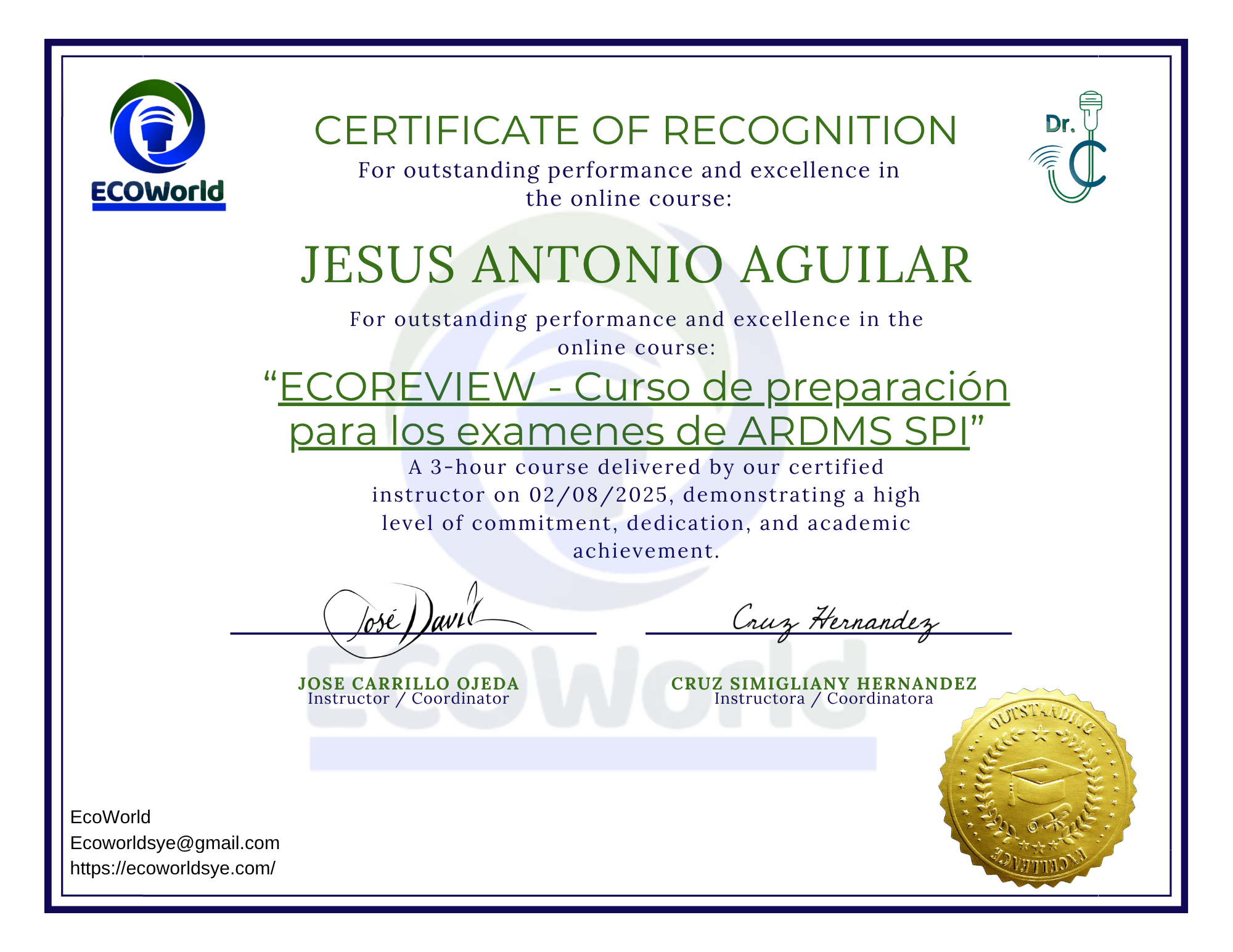
Certificado de finalización
Aplica a un certificado al completar el 80% de las lecciones vistas.
Certificado de finalización
Título del curso
$600.00
$900.00
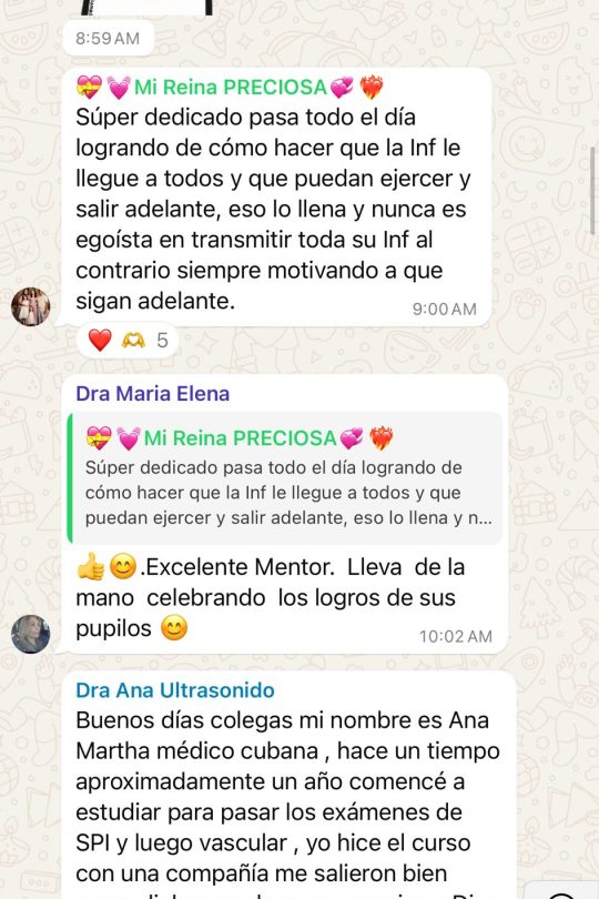
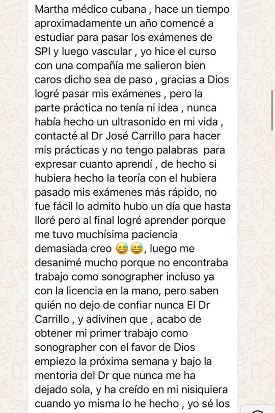
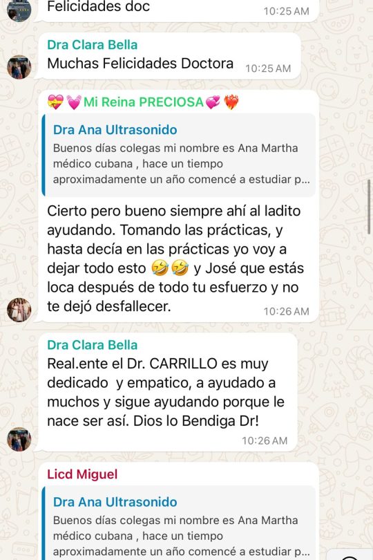
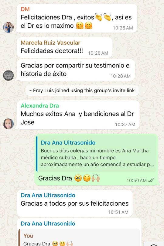
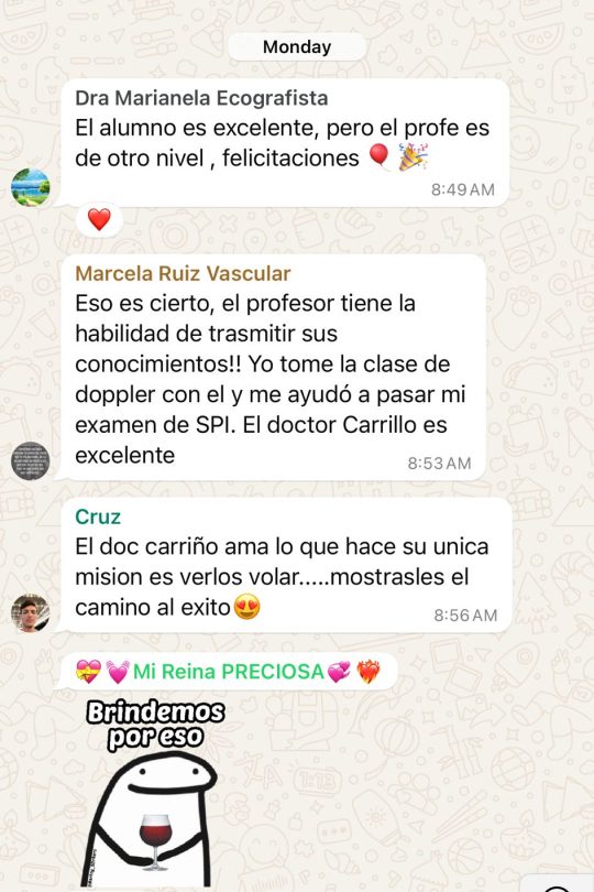
Nuestros estudiantes dicen sobre nosotros